Overview
Smarter Diagnostics, Powered by AI
PrediX delivers advanced, AI-driven solutions for radiology and pathology, helping health care professionals detect critical findings faster, reduce workload, and support more confident decision-making.
Modules
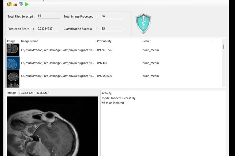
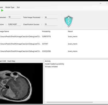
Radiology – MRI Triage & ROI Guidance
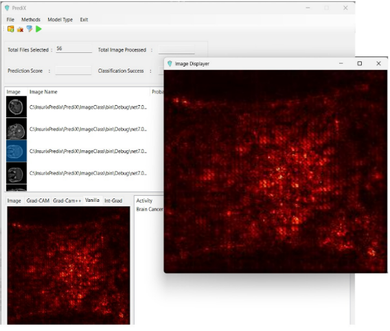
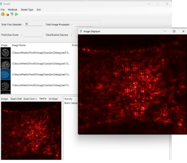
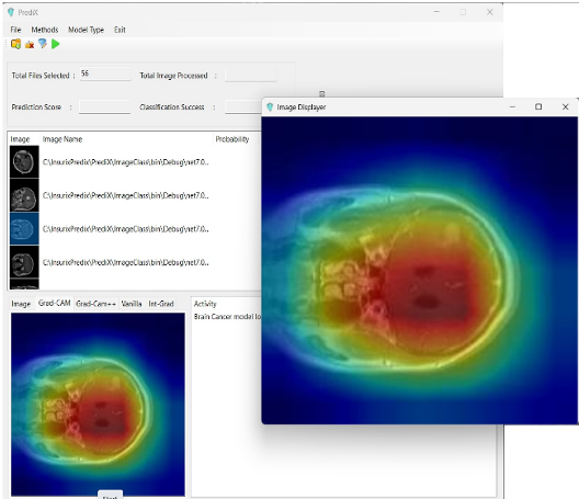
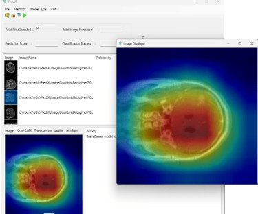
Our radiology module analyzes multi-sequence MRI scans to automatically highlight regions of interest, generate risk scores, and provide structured findings.
Inputs: Classified images folders
Outputs: Tumor probability of image and tumor classification
Benefits:
Accelerates triage and prioritization
Reduces radiologist workload
Enhances early and accurate decision-making
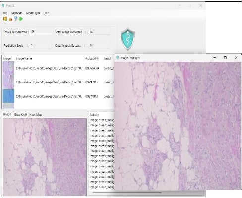
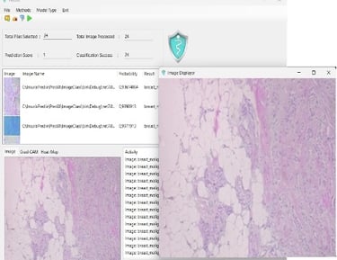
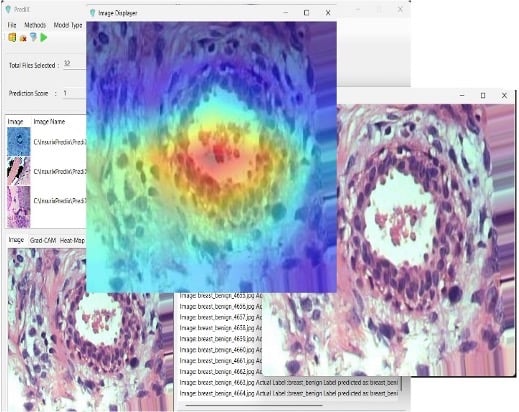
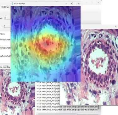
Pathology – WSI Region Proposals & Second-Read
AI-powered support for digital pathology.
The pathology module identifies critical diagnostic regions in whole slide images and provides second-read assistance to pathologists.Inputs: Classified images folders
Outputs: Tumor probability of image and tumor classification
Benefits:
Focuses attention on key regions
Improves diagnostic accuracy and efficiency
Fully compatible with leading digital pathology platforms
Intended Use
Decision support for trained clinicians; not a replacement for clinical judgment.
Clinical Accuracy
+90% accuracy; ongoing trials for lung, prostate, etc.
Integration Features
“PrediX enables Canadian hospitals to accelerate oncology diagnosis and treatment planning through AI-driven image classification. The platform is cloud-ready, PHIPA-compliant, and adaptable for integration into hospital IT systems.”
Current functionality: accepts standard image files (JPG, BMP, PNG, TIFF) in classified folders to train and predict new cases.
Optional integration modules (developed per site needs):DICOM / DICOMweb, HL7 v2, FHIR R4, SSO (SAML / OIDC), PACS / VNA connectors
PrediX can be configured to connect with PACS, VNA, or EHR systems based on client requirements.
Standard healthcare protocols (DICOM/DICOMweb, HL7 v2, FHIR R4, SSO via SAML/OIDC) are available as optional modules, to be developed per hospital platform.
Current production release supports direct image file input (JPG, BMP, PNG, TIFF) via structured folders (e.g., pituitary, pineal, healthy).
Flexible architecture allows customization for integration into each hospital’s IT environment.
Workflow Tools
Real-time AI insights
Automated reporting
Customizable dashboards
Training simulations
Safety
Human-in-the-loop, confidence bands, OOD detection, versioned models, audit trails.
Key capabilities
AI analysis for MRI and histopathology images
Real-time region-of-interest feedback
Adaptive learning with a simulation UI
Simple desktop and REST API integration for hospitals and enterprises
Primary outcomes we target
Fewer missed early-stage tumors
More consistent interpretation between readers
Faster turnaround under time pressure
Radiology AI (MRI)
What it does ?
PrediX applies convolutional neural networks to MRI studies to surface suspicious patterns and highlight regions for review, delivering immediate feedback as part of your reading workflow.
Highlights
CNN-based MRI analysis with real-time feedback
Validated on a large internal dataset (15,000+ MRI scans)
Desktop app or API-driven deployment options
Pathology AI (Histopathology)
What it does ?
For digital pathology slides, PrediX flags areas of interest and supports consistent grading and review, complementing your pathologists’ expertise and supporting second-look workflows.